

Nanomaterial-Based Sensor and Biosensor Development Services
The detection of disease biomarkers at clinically relevant concentrations presents one of the most demanding analytical challenges in modern diagnostics. A cancer biomarker present at femtogram-per-milliliter levels in blood, a pathogen circulating at single-copy-per-milliliter abundance, or a neurotoxin acting at parts-per-trillion concentrations: each demands sensing technology that operates at the physical limits of detection. Conventional analytical methods such as ELISA, PCR, and mass spectrometry achieve the required sensitivity but demand centralized laboratories, trained operators, and hours to days of processing time.
Nanomaterial-based sensors offer a fundamentally different approach. By exploiting the unique electronic, optical, and catalytic properties of nanoscale materials, these devices transduce molecular recognition events directly into measurable electrical or optical signals without amplification, labeling, or complex sample preparation. At Eata Nanomaterials, we design, fabricate, and validate nanosensor platforms that bridge the gap between research-grade sensitivity and practical diagnostic utility, delivering sensing systems capable of detecting target analytes at zeptomolar concentrations in complex biological matrices.
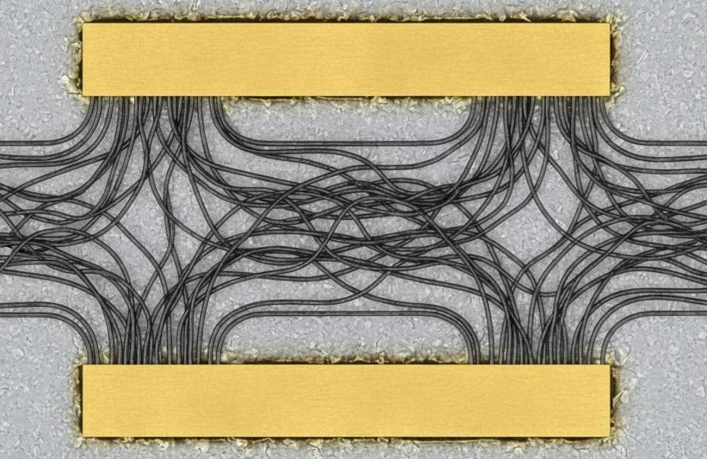 Figure 1: A carbon nanotube field-effect transistor biosensor chip showing semiconducting single-walled carbon nanotubes bridging between gold source and drain electrodes on a silicon dioxide surface.
Carbon Nanotube and Graphene Field-Effect Transistor Biosensors
Field-effect transistor biosensors based on one-dimensional carbon nanotubes and two-dimensional graphene represent the forefront of label-free electronic biosensing. In these devices, the nanocarbon channel serves as both the transduction element and the sensing surface. When a charged biomolecule binds to a receptor functionalized on the channel surface, the local electrostatic environment shifts, modulating the channel conductance in real time. This direct electrical readout eliminates the need for fluorescent labels, enzymatic amplification, or optical instrumentation.
Our CNT-FET and GFET biosensor development services encompass:
- Semiconducting carbon nanotube network fabrication by dielectrophoretic alignment or random network deposition onto silicon substrates with thermally grown oxide. We sort metallic and semiconducting nanotubes by density gradient ultracentrifugation or polymer wrapping to achieve semiconducting purities above 99 percent, ensuring high on-off ratios and reproducible transistor characteristics
- Graphene transfer and patterning by chemical vapor deposition growth on copper foils, followed by polymethyl methacrylate-assisted transfer to target substrates and lithographic patterning into device arrays. Monolayer quality is verified by Raman spectroscopy and optical contrast measurement before device fabrication
- Surface functionalization with biological recognition elements including antibodies, aptamers, DNA probes, and peptide ligands. Linker chemistry is selected for each application: 1-pyrenebutanoic acid succinimidyl ester for non-covalent attachment preserving graphene electronic properties, or silane and poly-L-lysine coupling for robust covalent immobilization
- Enlarged gate architectures and floating-gate designs that increase the effective sensing area and enhance gating efficiency, overcoming the Debye screening limitation that typically restricts FET biosensor performance in high-ionic-strength physiological samples
Demonstrated detection limits from our platform include osteoarthritis biomarker CRTAC1 at 0.2 femtograms per milliliter, phosphorylated tau-217 protein for Alzheimer disease diagnosis, lung cancer biomarkers, and SARS-CoV-2 at the single-virus level. Detection is achieved within minutes directly in undiluted serum or saliva, without sample pretreatment.
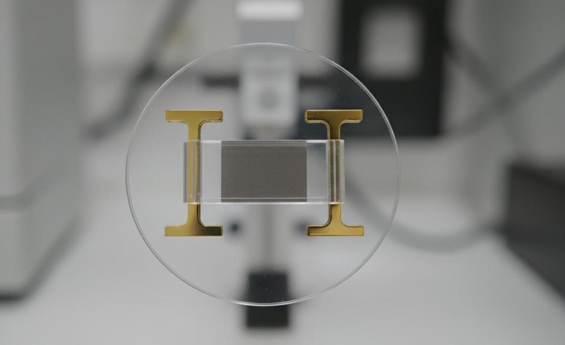 Figure 2: A graphene field-effect transistor biosensor device with a single-layer graphene channel between chromium-gold electrodes on a silicon substrate, with an integrated microfluidic delivery channel.
Surface-Enhanced Raman Scattering Sensor Platforms
Surface-enhanced Raman scattering amplifies the inherently weak Raman signals of molecules by factors of 10^8 to 10^10 when those molecules adsorb onto plasmonic nanostructures. This enhancement arises from localized electromagnetic field concentration at nanogaps and sharp features, combined with chemical enhancement through charge transfer between molecule and metal. The result is a vibrational fingerprint spectrum that simultaneously identifies and quantifies target molecules with single-molecule sensitivity.
Eata Nanomaterials fabricates SERS substrates and nanoparticle tags for diverse sensing applications:
- Gold nanoparticle aggregate substrates with controlled interparticle spacing for creating electromagnetic hot spots. We tune particle size, shape, and assembly conditions to optimize the enhancement factor for specific target molecules, achieving reproducible signals with relative standard deviations below 10 percent across substrate batches
- Core-shell nanoparticle designs including gold-silica, gold-polymer, and gold-magnetic configurations that combine SERS activity with additional functionality such as target enrichment, separation capability, or multimodal imaging contrast
- Machine learning-enhanced spectral analysis pipelines that deconvolute complex SERS spectra in mixtures, enabling multiplexed detection of multiple analytes from a single measurement. Statistical and artificial intelligence approaches address challenges that synthetic improvements alone cannot fully resolve
- Quantitative SERS protocols with rigorous internal standards and calibration methodologies, moving SERS from a qualitative technique to a reliable quantitative analytical tool suitable for clinical diagnostics and environmental monitoring
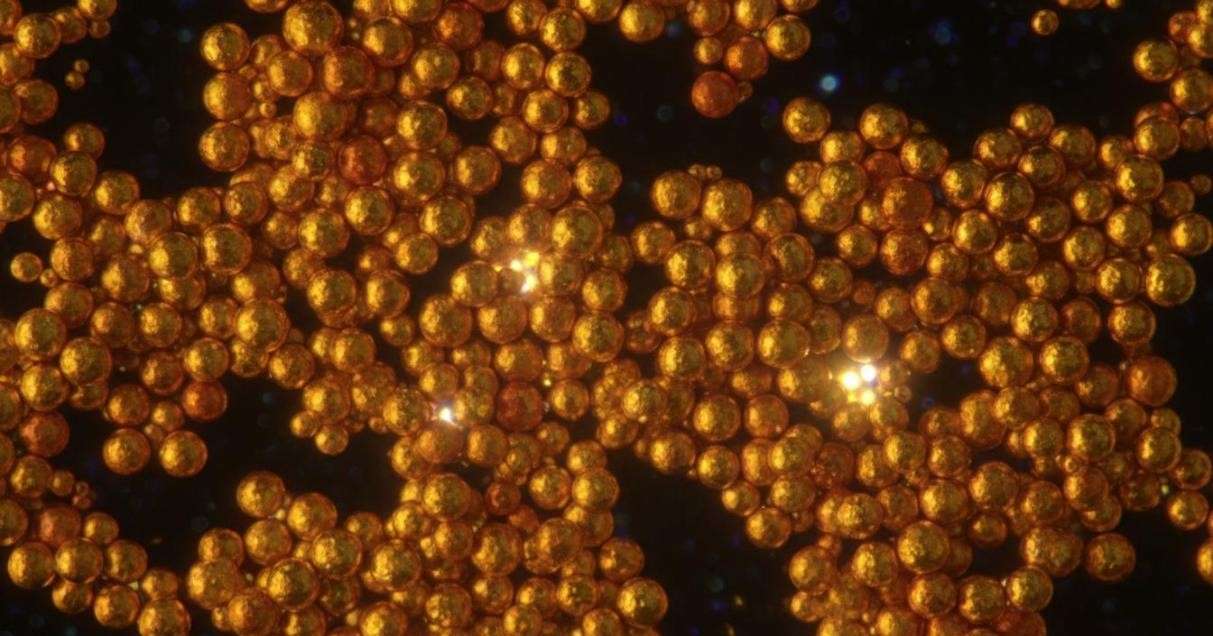 Figure 3: Aggregated gold nanoparticles forming plasmonic hot spots for surface-enhanced Raman scattering, with bright spots indicating localized electromagnetic field enhancement at particle junctions.
Quantum Dot Fluorescence Biosensors
Quantum dots possess extraordinary fluorescence properties that make them superior to organic fluorophores for biosensing: broad absorption spectra enabling excitation at any wavelength shorter than the emission peak, narrow and symmetric emission bands enabling multiplexed detection of multiple targets, exceptional photostability resisting photobleaching under continuous excitation, and size-tunable emission spanning the ultraviolet to near-infrared range.
Our quantum dot biosensor development capabilities include:
- Fluorescence resonance energy transfer assays where quantum dot donors are paired with organic dye or gold nanoparticle acceptors through molecular linkers that are cleaved or conformationally altered upon target binding. This architecture achieves ratiometric detection that is self-referencing against environmental fluctuations
- Sandwich fluorescence-linked immunosorbent assays utilizing quantum dot-labeled detection antibodies for ultra-sensitive detection of protein biomarkers, viral antigens, and bacterial toxins. The brightness of quantum dots enables detection limits 10 to 100-fold lower than conventional ELISA
- Molecularly imprinted polymer-quantum dot composites that combine the selectivity of artificial antibody-like binding cavities with the sensitivity of quantum dot fluorescence. These systems demonstrate remarkable selectivity with imprinting factors exceeding 6.7 and have been validated for antibiotic detection in serum with recoveries from 90 to 97 percent
- Near-infrared emitting quantum dots for deep-tissue imaging and in vivo biosensing applications where visible light cannot penetrate. PbS and Ag2S quantum dots emit in the 900 to 1600 nanometer range, enabling detection through several millimeters of biological tissue
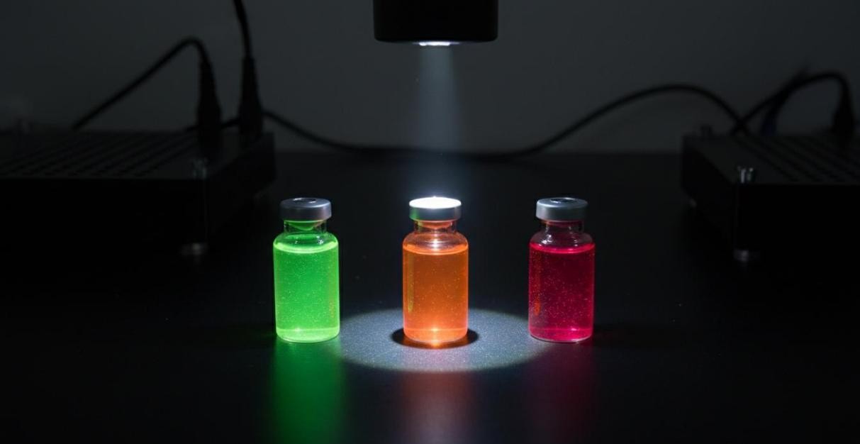 Figure 4: Three vials of colloidal quantum dots emitting green, orange, and red fluorescence under UV illumination, demonstrating size-tunable emission for multiplexed biosensing.
Electrochemical Sensor Development
Electrochemical sensors transduce chemical information into electrical signals through redox reactions at electrode surfaces. Nanomaterial integration dramatically enhances sensitivity by increasing electroactive surface area, accelerating electron transfer kinetics, and enabling catalytic amplification of target signals.
Eata Nanomaterials develops nanomaterial-enhanced electrochemical sensors for:
- Glucose and metabolite detection using gold nanoparticle-modified electrodes with immobilized oxidase enzymes. The nanoparticles improve enzyme loading, electron transfer efficiency, and electrode stability, achieving clinically relevant detection ranges with high selectivity against interfering species
- Heavy metal ion detection using bismuth film electrodes, mercury-free stripping voltammetry, and functionalized gold nanoparticles that selectively preconcentrate target ions. Detection limits reach parts-per-trillion levels for lead, cadmium, mercury, and arsenic in water and biological samples
- Neurotransmitter and drug monitoring with carbon nanotube and graphene-modified electrodes that resolve the overlapping voltammetric signals of dopamine, serotonin, and ascorbic acid through catalytic selectivity, enabling direct measurement in undiluted biological fluids
- Nucleic acid detection through electrochemical impedance spectroscopy at gold electrodes functionalized with capture probes, achieving femtomolar sensitivity for pathogen DNA and microRNA biomarkers without amplification
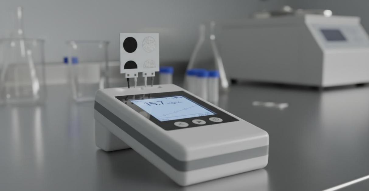 Figure 5: A portable electrochemical sensor device with a digital display and a disposable screen-printed electrode strip for point-of-care diagnostics in clinical or field settings.
Sensor Platform Comparison
| Platform | Transduction | Typical LOD | Key Advantage | Best For |
| CNT-FET | Electrical conductance | 0.2 fg/mL | Label-free, Real-time | Protein biomarkers |
| GFET | Field effect | zeptomolar | High mobility | Pathogen detection |
| SERS | Raman scattering | Single molecule | Molecular fingerprint | Chemical ID |
| QD-FRET | Fluorescence | pM range | Multiplexing | Immunoassays |
| Electrochemical | Redox current | ppb-ppt | Low cost, Portable | Metals, metabolites |
| QD-MIP | Fluorescence quenching | 50 ng/mL | High selectivity | Small molecules |
Application Areas
Our nanomaterial-based sensor development services support diverse diagnostic and analytical research programs:
- Oncology diagnostics: detecting circulating tumor markers, exosomal microRNAs, and cancer-associated proteins at concentrations far below conventional ELISA limits, enabling early-stage cancer screening from blood or saliva samples
- Neurodegenerative disease monitoring: quantifying phosphorylated tau proteins, amyloid-beta oligomers, and alpha-synuclein aggregates for Alzheimer and Parkinson disease diagnosis and progression tracking
- Infectious disease detection: rapid identification of bacterial pathogens, viral particles, and endotoxins at the single-copy or single-particle level, with results in under 15 minutes from sample to answer
- Environmental monitoring: detecting heavy metals, pesticides, and persistent organic pollutants in water and soil at regulatory-relevant concentrations using portable field-deployable sensor systems
- Food safety testing: screening for mycotoxins, allergens, and bacterial contamination in food products with sensitivity exceeding that of laboratory-based reference methods
Sensor Fabrication and Validation
Every sensor development project at Eata Nanomaterials follows a rigorous validation pipeline to ensure that demonstrated performance translates from buffer solutions to real-world samples. The process begins with receptor selection and surface chemistry optimization on test substrates, followed by device fabrication and electrical or optical characterization. Analytical figures of merit are determined: limit of detection, linear dynamic range, sensitivity, selectivity against interfering species, and reproducibility across device batches.
Clinical validation employs spiked samples and clinical specimens collected under appropriate ethical approvals. We assess matrix effects from serum, plasma, saliva, urine, and tissue lysates, and develop calibration strategies that compensate for sample-to-sample variability. Long-term stability studies evaluate sensor shelf life under various storage conditions, and accelerated aging protocols predict device lifetime for commercialization planning.
If you are interested in our products or services, please don't hesitate to contact us.