

Custom Nanoparticle Synthesis Services
Nanoparticles are the building blocks of modern nanomedicine, diagnostics, and advanced materials science. Yet off-the-shelf nanoparticles rarely meet the exacting requirements of cutting-edge research. Particle size, surface chemistry, morphology, dispersibility, and batch consistency must all be precisely controlled to achieve reproducible results. At Eata Nanomaterials, we specialize in synthesizing bespoke nanoparticle systems engineered to your exact specifications, supported by comprehensive characterization and consultation at every stage.
Our synthesis platform covers metallic, metal oxide, semiconductor, silica, carbon-based, and polymeric nanoparticles across a broad size range — from sub-5 nm quantum dots to 500 nm mesoporous carriers. Whether you require a small research batch for proof-of-concept studies or larger quantities for preclinical evaluation, our chemists deliver nanoparticles with precisely defined critical quality attributes that accelerate your research and eliminate the uncertainty of in-house synthesis.
Noble Metal Nanoparticle Synthesis
 Figure 1: Colloidal gold nanoparticles synthesized via citrate reduction showing characteristic ruby-red color and uniform spherical morphology
Gold and silver nanoparticles have earned a central place in nanotechnology due to their unique optical properties — surface plasmon resonance generates intense absorption and scattering in the visible and near-infrared range — combined with exceptional biocompatibility and versatile surface chemistry. Our noble metal nanoparticle synthesis service produces monodisperse colloids with tunable sizes and shapes, enabling applications in biosensing, photothermal therapy, diagnostic imaging, and as contrast-enhancing agents.
Beyond conventional spherical particles, we synthesize anisotropic morphologies including gold nanorods, nanoshells, nanostars, and nanocages. Each shape exhibits distinct plasmonic properties: nanorods offer tunable longitudinal absorption bands in the NIR tissue transparency window, while nanostars generate intense electromagnetic field enhancements at their tips for surface-enhanced Raman scattering applications.
Synthesis capabilities at a glance:
- Gold nanoparticles (spheres): Citrate reduction, Brust-Schiffrin, and seed-mediated growth methods; diameters from 3 nm to 200 nm with coefficient of variation <10%.
- Gold nanorods: Seed-mediated surfactant template synthesis with aspect ratios from 2 to 20, tunable longitudinal surface plasmon resonance from 650 nm to 1100 nm.
- Gold nanoshells & nanocages: Galvanic replacement and sacrificial template methods for hollow structures with tunable plasmonic absorption optimized for photothermal applications.
- Silver nanoparticles: Chemical reduction with controlled nucleation and growth; sizes 5-100 nm with options for spherical, cubic, and triangular morphologies.
- Bimetallic nanoparticles: Au-Ag core-shell, alloy, and heterogeneous dimer structures combining the catalytic and optical properties of both metals.
Metal Oxide & Inorganic Nanoparticle Synthesis
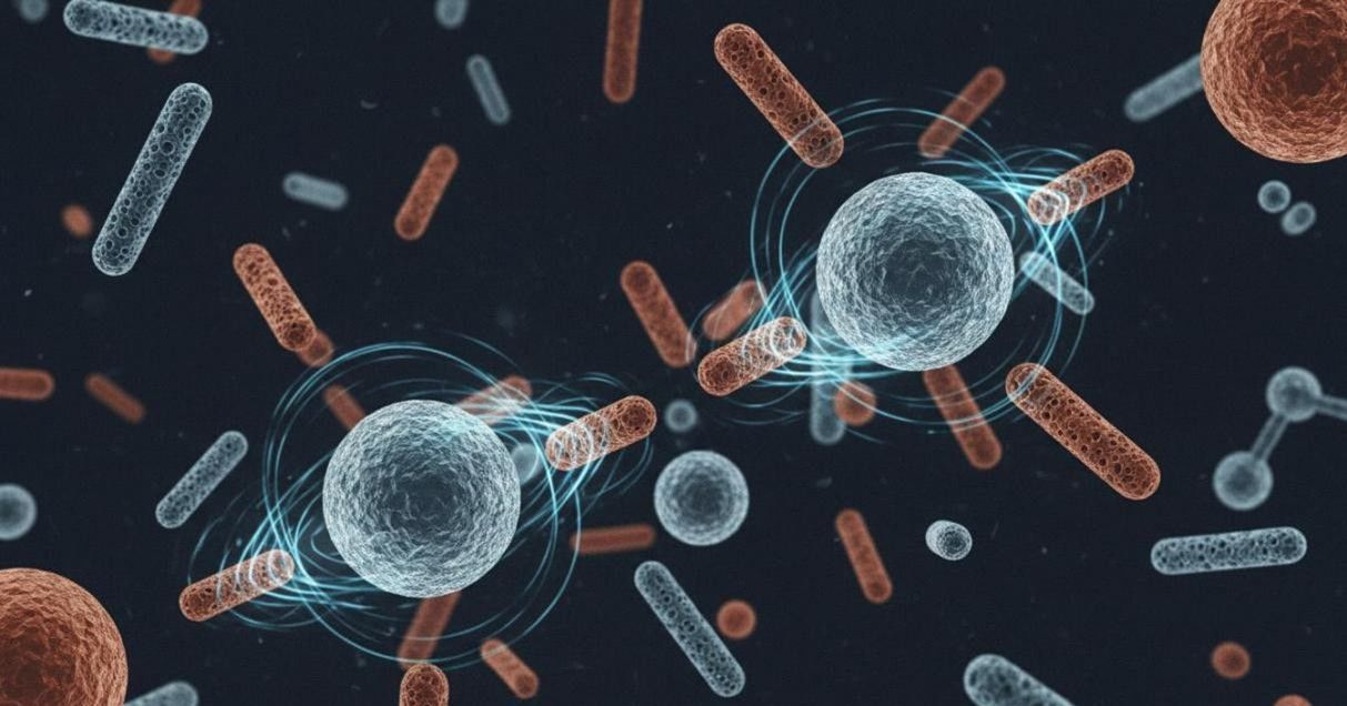 Figure 2: Metal oxide nanoparticles including magnetite spheres and mesoporous silica rods with magnetic field interaction visualization
Magnetic, semiconducting, and porous metal oxide nanoparticles serve critical roles across imaging, separation, catalysis, and drug delivery. Our metal oxide synthesis program leverages sol-gel chemistry, thermal decomposition, hydrothermal synthesis, and co-precipitation methods to produce particles with precisely controlled crystallinity, size, and surface functionality.
Iron oxide nanoparticles — particularly magnetite (Fe3O4) and maghemite (gamma-Fe2O3) — are the cornerstone of magnetic resonance imaging contrast enhancement, magnetic hyperthermia, and magnetic cell separation. We synthesize superparamagnetic iron oxide nanoparticles (SPIONs) across a size range from 5 nm to 50 nm with tight size distributions, high saturation magnetization, and surface coatings optimized for aqueous stability. Mesoporous silica nanoparticles, meanwhile, offer enormous surface area and precisely engineered pore structures ideal for high-capacity drug loading and controlled release.
| Material | Key Properties | Applications |
| Fe3O4 / gamma-Fe2O3 | Superparamagnetic, high Ms, T2 MRI contrast | MRI contrast, magnetic separation, hyperthermia |
| Mesoporous SiO2 | High surface area (>700 m2/g), tunable pores 2-10 nm | Drug delivery, catalysis, molecular sieving |
| TiO2 | Photocatalytic, UV absorption, biocompatible | Antimicrobial coatings, photocatalysis, sunscreens |
| ZnO | UV emission, antibacterial, piezoelectric | Wound healing, UV protection, sensors |
| CeO2 | Catalytic redox cycling, antioxidant mimicry | Anti-inflammatory therapy, catalysis, polishing |
| Al2O3 | High hardness, chemical stability, bioinert | Implant coatings, abrasive, chromatography |
Semiconductor & Carbon-Based Nanoparticle Synthesis
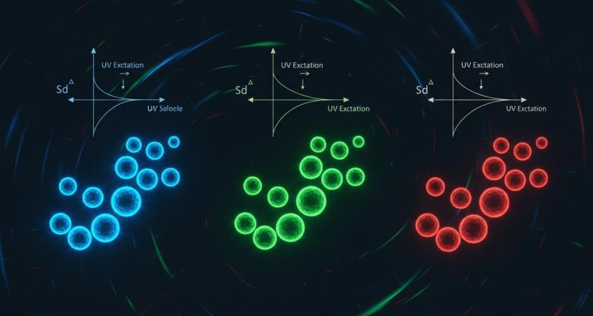 Figure 3: Size-tunable quantum dots exhibiting blue, green, and red fluorescence emission under UV excitation
Quantum dots, carbon dots, and graphene-based nanomaterials provide extraordinary optical and electronic properties that traditional fluorophores and bulk materials cannot match. Our semiconductor and carbon nanoparticle synthesis services deliver precisely engineered nanocrystals with tunable fluorescence, high quantum yield, and surface chemistry ready for bioconjugation, imaging, and sensing applications.
Platforms we synthesize:
- CdSe/ZnS core-shell quantum dots: Hot-injection organometallic synthesis with tunable emission from blue to deep red; high quantum yield (>70%) and excellent photostability.
- InP/ZnS quantum dots: Cadmium-free alternative with emission tunable across the visible spectrum for applications requiring reduced heavy metal content.
- Carbon dots & graphene quantum dots: Hydrothermal carbonization of organic precursors; excitation-dependent emission, low toxicity, and water solubility.
- Upconversion nanoparticles: NaYF4:Yb,Er/Tm hexagonal-phase nanocrystals that convert NIR excitation to visible emission for deep-tissue imaging.
- Single-wall & multi-wall carbon nanotubes: CVD synthesis with controlled diameter and chirality; functionalized for enhanced dispersibility and biocompatibility.
Polymeric & Organic Nanoparticle Synthesis
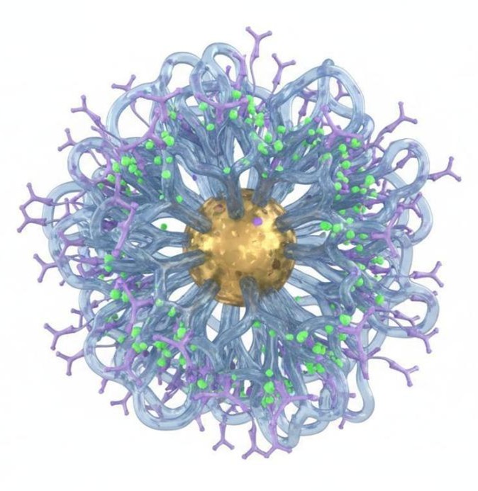 Figure 4: Multifunctional nanoparticle showing gold core with PEG polymer coating, antibody ligands, and fluorescent dye surface modification
Polymeric nanoparticles offer unmatched flexibility in drug loading, release kinetics, and surface functionalization. Our polymeric nanoparticle synthesis service produces biodegradable and biocompatible carriers through well-controlled emulsion, nanoprecipitation, and microfluidic methods, yielding particles with narrow size distributions, high encapsulation efficiency, and tailorable degradation profiles.
We synthesize polymeric nanoparticles from a wide range of FDA-approved and research-grade materials. PLGA and PLA particles remain the most widely used platforms for controlled drug release due to their excellent safety record and tunable degradation. For more specialized applications, we work with PCL, chitosan, albumin, gelatin, and a variety of diblock and triblock copolymers that self-assemble into micelles and polymersomes.
Polymer systems and methods:
- PLGA / PLA nanoparticles: Emulsion-solvent evaporation or nanoprecipitation; sizes 50-500 nm, tunable drug loading and sustained release from days to months.
- Block copolymer micelles: Self-assembly of PEG-PLA, PEG-PCL, and PEG-PLGA amphiphiles; 20-100 nm core-shell structures for solubilizing hydrophobic drugs.
- Polymersomes: Vesicular structures formed by high molecular weight block copolymers; robust bilayer membranes with enhanced stability over liposomes.
- Protein nanoparticles: Albumin, gelatin, and ferritin-based particles with natural biocompatibility and intrinsic targeting ligands.
- Hydrogel nanoparticles (nanogels): Crosslinked polymeric networks with high water content; ideal for protein and nucleic acid encapsulation with stimuli-responsive release.
Precision Surface Functionalization Services
The surface of a nanoparticle is where it meets the biological world. Pristine as-synthesized nanoparticles rarely possess the surface chemistry required for specific applications. Our surface functionalization service transforms raw nanocrystals into purpose-built tools by introducing precisely defined chemical groups, polymer coatings, targeting ligands, and detection labels.
| Functionalization Type | Capabilities |
| Reactive groups | Carboxyl (-COOH), amine (-NH2), thiol (-SH), hydroxyl (-OH), aldehyde (-CHO), azide/alkyne click-chemistry handles |
| PEGylation | Linear and branched PEG (1-20 kDa) for stealth, improved circulation, and reduced immunogenicity |
| Targeting ligands | Antibodies, Fab fragments, peptides (RGD, TAT), aptamers, folate, transferrin, galactose for active targeting |
| Fluorescent labels | FITC, Cy3, Cy5, Alexa Fluor dyes, quantum dots for imaging and flow cytometry applications |
| Bio-orthogonal handles | DBCO, tetrazine, TCO, biotin for rapid, specific conjugation under mild conditions |
| Stimuli-responsive coatings | pH-sensitive, thermosensitive, and enzyme-cleavable coatings for smart, triggered behavior |
Core-Shell & Hybrid Nanostructure Engineering
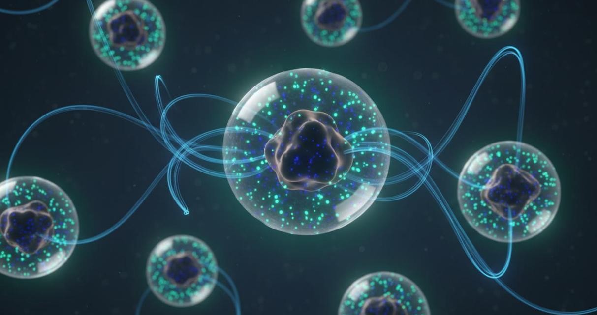 Figure 5: Core-shell magnetic-fluorescent nanoparticles with iron oxide core and dye-doped silica shell for dual-modal imaging
Combining multiple functional materials into a single nanostructure unlocks synergistic capabilities that individual components cannot achieve alone. Our core-shell and hybrid nanostructure engineering service designs and fabricates multi-functional particles that integrate magnetic, optical, therapeutic, and targeting functions within a unified architecture.
We fabricate core-shell structures through layer-by-layer deposition, sol-gel coating, emulsion templating, and seeded growth methods. Silica shells protect and stabilize sensitive cores while providing a versatile surface for functionalization. Polymer shells enable drug loading and controlled release. Metal shells introduce plasmonic or catalytic properties. The permutations are nearly limitless, and we work with you to identify the optimal architecture for your application.
Hybrid architectures we develop:
- Magnetic-fluorescent nanoparticles: Iron oxide core with fluorescent silica or polymer shell for dual MRI-optical imaging and magnetic manipulation.
- Plasmonic-magnetic nanostars: Gold nanostar-coated magnetic particles combining photothermal conversion with magnetic targeting capabilities.
- Upconversion-photosensitizer hybrids: NaYF4 core with rose bengal or chlorin e6-conjugated shell for NIR-activated photodynamic therapy.
- Janus nanoparticles: Bifacial particles with two distinct surface chemistries enabling simultaneous loading of incompatible payloads.
- MOF-nanoparticle composites: Metal-organic frameworks grown on nanoparticle seeds for gas storage, catalysis, and stimuli-responsive drug delivery.
Comprehensive Nanoparticle Characterization
Rigorous characterization is the cornerstone of reliable nanoparticle synthesis. Every batch we produce undergoes thorough physicochemical analysis to confirm that critical quality attributes meet specifications. Our characterization data packages support research publication, patent filing, and regulatory submission requirements.
- Size and morphology: Dynamic light scattering (DLS) for hydrodynamic diameter and polydispersity index; transmission electron microscopy (TEM) and scanning electron microscopy (SEM) for direct visualization of size, shape, and crystallinity.
- Surface properties: Zeta potential measurement for colloidal stability prediction; X-ray photoelectron spectroscopy (XPS) and Fourier-transform infrared spectroscopy (FTIR) for surface chemistry confirmation.
- Crystallographic analysis: X-ray diffraction (XRD) for phase identification, crystallite size determination, and lattice parameter calculation.
- Optical and magnetic properties: UV-Vis absorption and fluorescence spectroscopy for plasmonic and luminescent particles; vibrating sample magnetometry (VSM) and SQUID magnetometry for magnetic characterization.
- Elemental composition: Inductively coupled plasma mass spectrometry (ICP-MS) and energy-dispersive X-ray spectroscopy (EDX) for quantitative elemental analysis.
- Stability assessment: Accelerated aging studies, freeze-thaw cycling, and serum stability testing to evaluate colloidal stability under relevant storage and biological conditions.
Begin Your Custom Nanoparticle Project Today
Reach out to Eata Nanomaterials to discuss your specific nanoparticle requirements. Our synthetic chemists will evaluate your material, size, surface chemistry, and quantity needs to develop a tailored synthesis protocol that delivers precisely what your research demands.