

Crystallographic and Phase Analysis Services (XRD)
X-ray diffraction stands as one of the most powerful and widely employed techniques for characterizing the crystallographic structure of nanomaterials. When X-rays interact with a crystalline sample, the periodic arrangement of atoms produces constructive interference at specific angles, generating a diffraction pattern that serves as a unique structural fingerprint. This fingerprint reveals phase composition, crystallite dimensions, lattice parameters, and internal strain, information essential for correlating synthesis conditions with material performance.
At Eata Nanomaterials, our XRD laboratory is equipped with modern powder diffractometers operating with copper K-alpha radiation, offering comprehensive crystallographic analysis services from routine phase identification to advanced Rietveld refinement. Our analytical team combines deep expertise in diffraction physics with practical experience across diverse nanomaterial classes including metal oxides, quantum dots, perovskites, MXenes, MOFs, and two-dimensional materials.
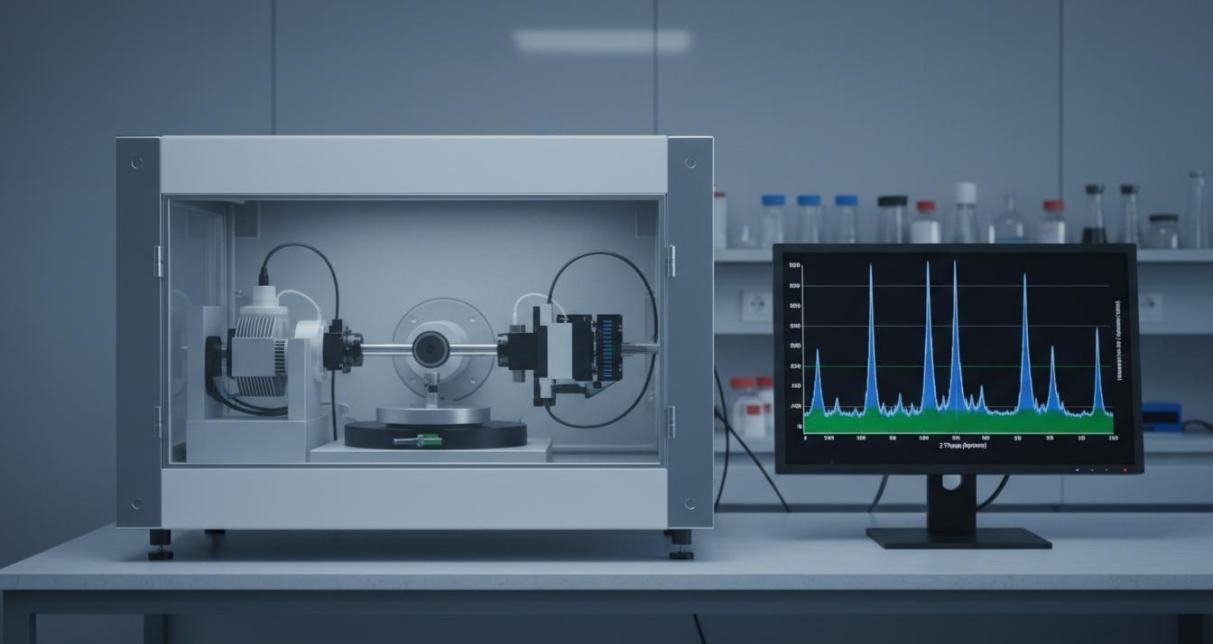 Figure 1: An X-ray diffractometer system with protective cabinet and workstation displaying a characteristic diffraction pattern.
Phase Identification and Purity Verification
Every crystalline material produces a diffraction pattern unique to its atomic arrangement. By comparing measured patterns against international crystallographic databases such as the ICDD PDF and COD, we identify the phases present in a sample with high confidence. This capability underpins quality control, synthesis optimization, and failure analysis across nanomaterial research programs.
Our phase identification services include:
- Qualitative phase analysis determining which crystalline phases are present in a sample, including major phases, minor impurities, and trace contaminants
- Quantitative phase analysis using Rietveld refinement or reference intensity ratio methods to calculate the weight fraction of each phase in multicomponent mixtures
- Polymorph screening distinguishing different crystal structures of the same chemical composition, such as anatase versus rutile in TiO2 nanoparticles
- Solid solution analysis tracking lattice parameter shifts that indicate dopant incorporation or compositional variation within a single phase
For nanomaterials, peak broadening complicates phase identification. Our analysts apply profile fitting algorithms that deconvolute overlapping peaks, and we employ instrumental standards to separate size-related broadening from true structural effects.
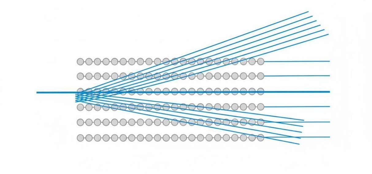 Figure 2: A schematic illustration of X-rays diffracting from parallel atomic crystal planes according to Bragg's law.
Crystallite Size and Microstrain Analysis
A critical distinction in nanomaterial science exists between particle size and crystallite size. A single nanoparticle may contain multiple crystalline domains separated by grain boundaries, or conversely, a large agglomerate may consist of a single crystallite. XRD uniquely probes the crystallite size through analysis of diffraction peak broadening, providing complementary information to electron microscopy and light scattering techniques.
We employ multiple approaches to extract crystallite size and microstrain from diffraction line profiles:
Scherrer equation: The classical approach relating peak broadening to crystallite dimensions through the formula L = K lambda / beta cos theta. We apply this method with shape factor K typically set to 0.89 for spherical crystals, selecting the most intense and well-resolved peak for rapid size estimation.
Modified Scherrer method: A least-squares refinement plotting ln(beta) against ln(1/cos theta) across all available reflections. This approach yields a single crystallite size value incorporating data from multiple peaks, significantly reducing errors associated with peak-specific broadening effects.
Williamson-Hall analysis: Separates size-induced broadening from strain-induced broadening by plotting beta cos theta versus sin theta. The intercept yields crystallite size while the slope provides the microstrain value, enabling simultaneous quantification of both structural parameters.
Rietveld refinement: Full-pattern fitting that models size and strain through profile shape functions. This method provides the most accurate results for complex patterns with overlapping peaks, simultaneously refining lattice parameters, occupancies, and thermal parameters.
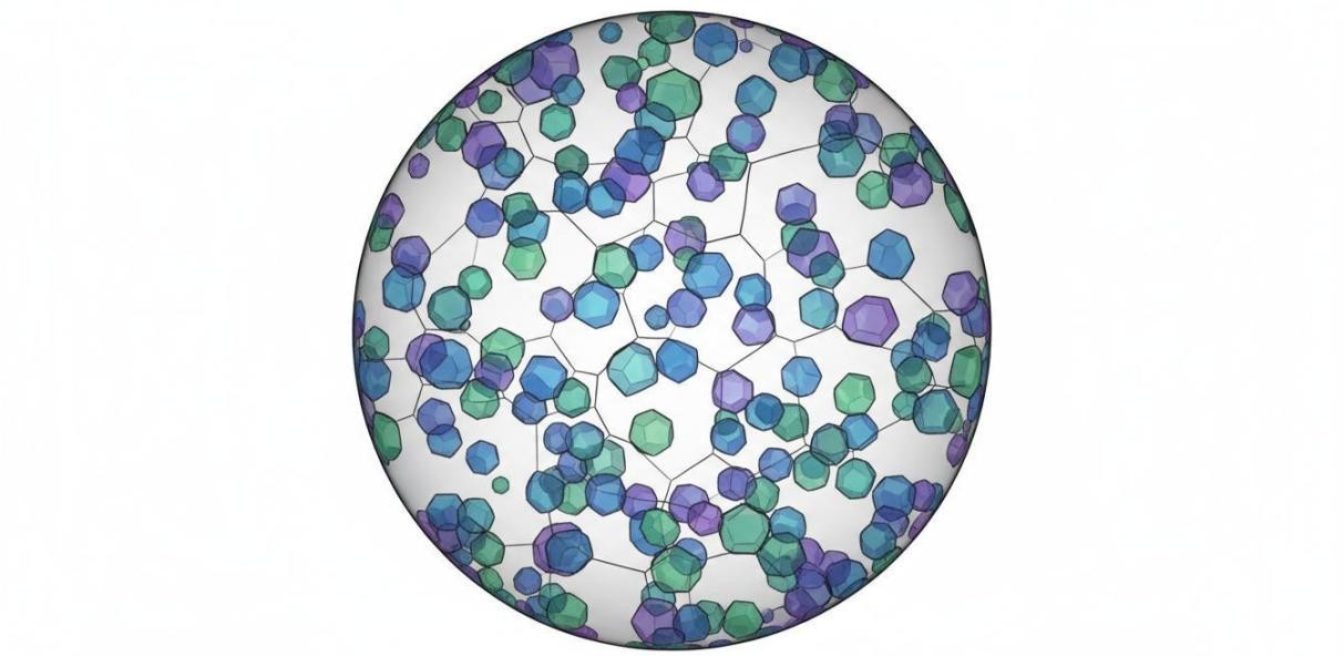 Figure 3: A spherical nanoparticle containing multiple crystalline domains with different orientations, illustrating the crystallite concept.
Rietveld Refinement for Comprehensive Structural Analysis
Rietveld refinement represents the gold standard for extracting detailed crystallographic information from powder diffraction data. By iteratively adjusting a structural model until the calculated pattern matches the experimental data, this method provides quantitative insights unattainable through simpler peak-by-peak analysis.
Our Rietveld refinement services deliver:
- Precise lattice parameters with uncertainties typically in the fourth decimal place for unit cell dimensions
- Atomic positions and site occupancies revealing structural distortions, defects, and non-stoichiometry
- Quantitative phase analysis with weight fractions for all crystalline components in multiphase samples
- Crystallite size and microstrain values through profile shape modeling
- Texture analysis quantifying preferred orientation effects common in thin films and nanoparticle assemblies
- Goodness-of-fit parameters including Rwp, Rexp, and chi-squared values documenting refinement quality
We support refinements across all major crystal systems and space groups, with particular expertise in perovskite oxides, metal oxide nanoparticles, battery electrode materials, and pharmaceutical polymorphs.
Thin Film and Coating Analysis by GIXRD
Conventional Bragg-Brentano geometry suffers significant limitations when analyzing thin films deposited on substrates, as the majority of the X-ray beam penetrates through the film and diffracts from the underlying bulk material. Grazing incidence X-ray diffraction (GIXRD) overcomes this limitation by fixing the incident X-ray beam at a shallow angle, concentrating the probe within the film itself.
Our GIXRD capabilities include:
- Phase identification of nanometer-scale films without substrate interference, with detection capability down to sub-100 nm thicknesses
- Depth profiling by varying the incident angle to probe successive layers from the surface downward, revealing composition gradients and interlayer phases
- Texture analysis measuring preferred orientation and grain alignment in epitaxial and polycrystalline films
- In-plane GIXRD for measuring lattice parameters and strain within the film plane, critical for understanding substrate-induced epitaxial strain
- Residual stress analysis through the multi-hkl method correlating peak shifts with stress magnitude and direction
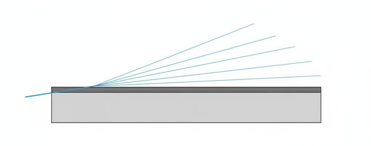 Figure 4: A grazing incidence X-ray diffraction setup showing the incident beam at shallow angle and diffracted beams from the thin film surface.
Temperature-Dependent and In-Situ Phase Evolution
Nanomaterial phases frequently transform during thermal processing, mechanical treatment, or chemical reaction. Our temperature-dependent XRD services track these structural evolutions in real time, providing kinetic and thermodynamic insights critical for process optimization.
Applications of temperature-dependent XRD in nanomaterial research include:
- Tracking crystallization kinetics of amorphous precursors to nanocrystalline products, identifying optimal annealing temperatures
- Monitoring phase transitions such as the anatase-to-rutile transformation in TiO2, or tetragonal-to-cubic transitions in perovskite systems
- Detecting thermal decomposition or oxidation of metastable phases that may compromise long-term material stability
- Quantifying thermal expansion through lattice parameter tracking as a function of temperature
Nanomaterial Classes We Characterize
| Material Class | Typical Information Obtained |
| Metal oxide nanoparticles | Phase purity, crystallite size, lattice parameters, dopant incorporation |
| Quantum dots | Crystal structure, size-dependent lattice contraction, shell composition |
| Perovskite nanocrystals | Phase purity, tolerance factor, halide ratio, thermal stability |
| MXenes | Interlayer spacing, termination group effects, delamination quality |
| MOFs / COFs | Crystallinity, pore structure, framework stability, guest inclusion |
| 2D materials | Layer stacking, intercalation, phase identification, defect density |
| Thin film coatings | Phase, texture, thickness, strain, depth-dependent composition |
| Battery materials | Phase transitions during cycling, lattice expansion, structural fatigue |
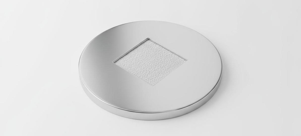 Figure 5: A metallic XRD sample holder with a fine powder specimen evenly spread across the measurement area.
Analysis Deliverables
Every XRD analysis project includes a comprehensive report with the following deliverables:
- Raw and processed diffraction data in standard file formats for independent analysis or database deposition
- Identified phases with ICDD/COD reference codes, confidence levels, and quantitative weight fractions
- Crystallite size and microstrain values with calculated uncertainties and methodology documentation
- Lattice parameters with standard deviations for each identified phase
- Publication-quality figures showing measured patterns, fitted profiles, and difference curves
- Expert interpretation addressing your specific research questions and recommending follow-up analyses when appropriate
Submit Your Samples for XRD Analysis
Whether you need rapid phase confirmation for a single batch, comprehensive Rietveld refinement for publication, or systematic temperature-dependent studies for process development, Eata Nanomaterials provides precise, reliable XRD characterization backed by decades of collective expertise in nanomaterial crystallography.
Contact our analytical team to discuss your sample type, desired measurements, and reporting requirements. We will recommend the optimal analytical approach and provide guidance on sample quantity, preparation, and shipping.