

Membrane-Coated Biomimetic Nanostructures Customized Services
Nature has spent billions of years engineering the perfect delivery vehicles: living cells. Erythrocytes circulate for months without triggering immune surveillance. Platelets instinctively home to sites of vascular injury. Cancer cells display an uncanny ability to recognize their own kind. At Eata Nanomaterials, we capture these innate biological advantages by coating functional nanocores with natural cell membranes, creating biomimetic nanostructures that masquerade as the body's own cells while carrying therapeutic payloads to precisely where they are needed.
Our membrane-coated biomimetic nanostructures merge the engineerable versatility of synthetic nanoparticles — tunable size, high loading capacity, controllable release — with the complex, multivalent surface biology of living cells. The resulting hybrid systems evade immune clearance, prolong circulation, and actively target diseased tissues in ways that conventional PEGylated nanoparticles simply cannot match. From concept to characterization, we build these sophisticated delivery platforms according to your exact therapeutic requirements.
Biomimetic Membrane Coating Technology Platform
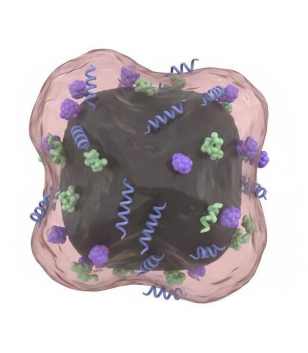 Figure 1: Red blood cell membrane-coated nanoparticle with phospholipid bilayer and embedded surface proteins
The heart of our service is the precise and reproducible coating of nanoparticle cores with intact, cell-derived membrane vesicles. Unlike synthetic surface modifications that introduce one ligand at a time, cell membrane coatings transfer the entire surface proteome, lipidome, and glycome of the source cell onto the nanocarrier — a feat of molecular mimicry that imparts multiple functions simultaneously.
We have refined the entire membrane harvesting and coating workflow to yield uniform, fully encapsulated nanostructures with preserved membrane orientation and protein functionality. Our platform accommodates diverse nanocore materials including polymeric nanoparticles (PLGA, PLA, PCL), mesoporous silica, metal-organic frameworks, gold nanoparticles, iron oxide nanocrystals, and upconversion nanomaterials.
Core capabilities of our membrane coating platform:
- Membrane extraction & vesicle preparation: Hypotonic lysis, freeze-thaw cycling, and sucrose density gradient ultracentrifugation to obtain intact membrane fragments that retain native protein orientation and activity.
- Membrane-protein quantification: BCA assay and Western blot validation to ensure consistent protein loading across batches — a critical quality attribute for reproducibility.
- Coating process optimization: Systematic tuning of membrane-to-core mass ratio, sonication energy, extrusion parameters, and incubation conditions to achieve complete, uniform coating without aggregation.
- Orientation verification: Confirmation that membrane proteins maintain their natural right-side-out orientation through immunogold labeling and flow cytometry.
- Scalability: Process development for batch sizes ranging from milligram research quantities to gram-scale preclinical production.
Cell Membrane Source Selection & Customization
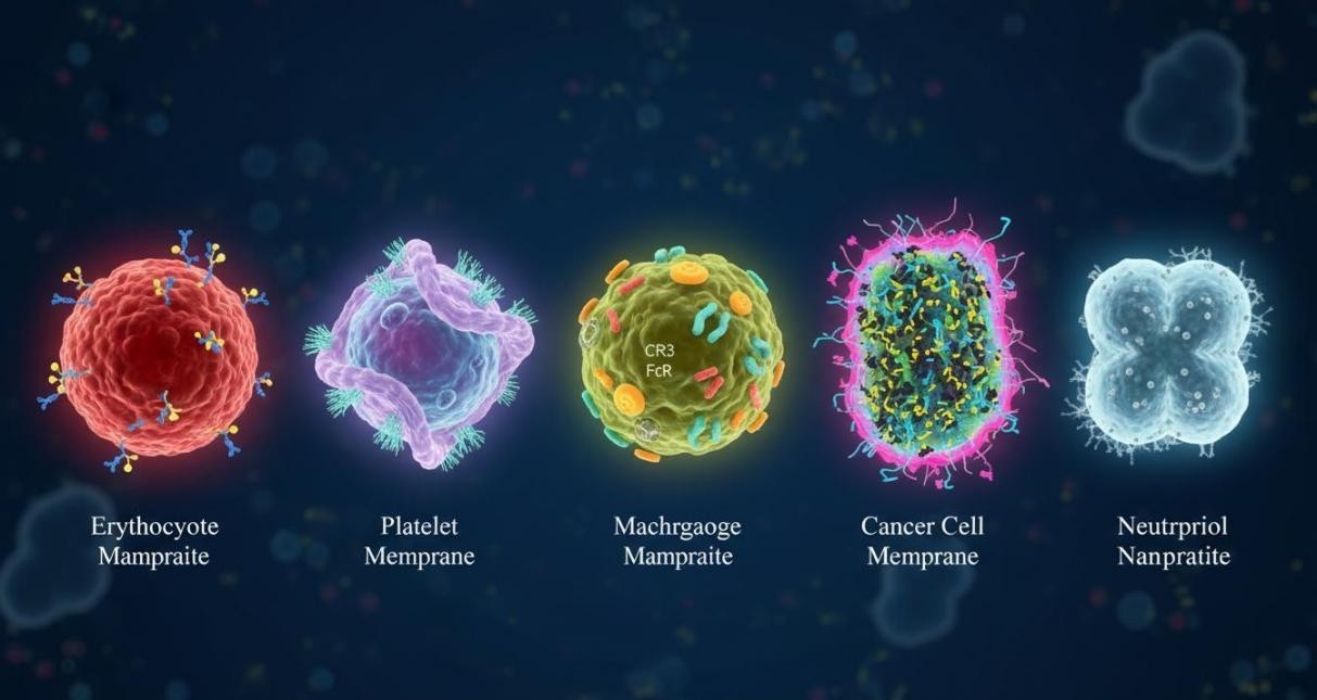 Figure 2: Diversity of cell membrane sources for biomimetic nanostructure engineering — erythrocyte, platelet, macrophage, cancer cell, and neutrophil membranes
The choice of membrane donor cell determines the biological identity and targeting behavior of the resulting nanostructure. We offer a comprehensive portfolio of membrane sources, each conferring distinct functional advantages for specific therapeutic scenarios. Our team guides you through selection based on your target tissue, desired circulation profile, and required biological interactions.
| Membrane Source | Key Surface Markers & Functions | Therapeutic Applications |
| Erythrocyte (RBC) | CD47 ("don't eat me" signal); long-circulating; flexible membrane | Prolonged circulation, reduced RES clearance, passive tumor accumulation |
| Platelet | GPVI, CD41, P-selectin; adhesion to collagen, activated endothelium, CTCs | Vascular injury targeting, thrombosis, metastasis inhibition |
| Macrophage | Integrins, Fc receptors, cytokine receptors; inflammatory homing | Tumor-targeted delivery, atherosclerosis, inflammatory diseases |
| Neutrophil | CXCR4, CD11b, L-selectin; migrates to inflamed tissues | Pancreatic cancer, CTC capture, bacterial infection targeting |
| Cancer cell | Homotypic adhesion molecules (E-cadherin, N-cadherin, EpCAM); tumor antigens | Homotypic tumor targeting, metastasis inhibition, cancer vaccines |
| Dendritic cell | MHC I/II, CD80, CD86; potent antigen presentation | Cancer immunotherapy, vaccine adjuvant, T cell activation |
| Stem cell | Homing receptors, cytokine receptors; regenerative signaling | Tissue repair, regenerative medicine, ischemia targeting |
| Hybrid/fusion membranes | Combinations of two or more membrane types via membrane fusion | Dual-targeting, enhanced immune modulation, multifunctional delivery |
Therapeutic Nanocore Engineering
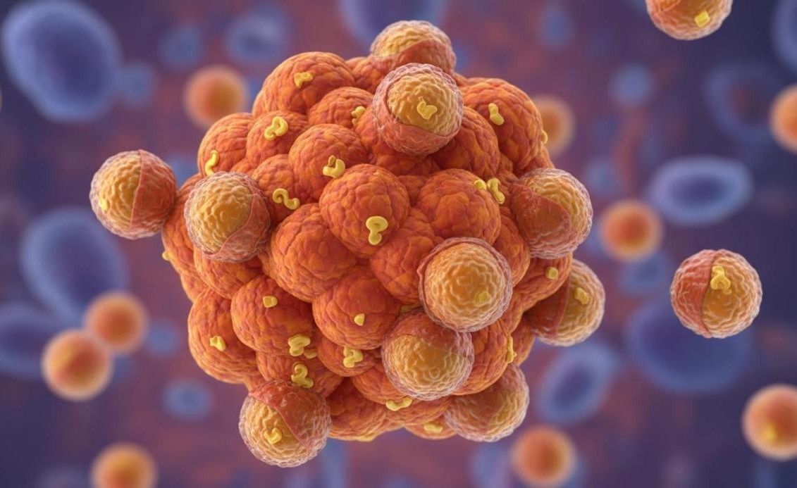 Figure 3: Cancer cell membrane-coated nanoparticles exhibiting homotypic targeting and accumulation within tumor tissue
The nanocore at the center of a membrane-coated biomimetic structure serves as the functional engine — providing payload capacity, release control, and additional therapeutic modalities. We engineer nanocores with precise control over size, morphology, porosity, and surface chemistry to complement the biological functions conferred by the membrane coating.
Nanocore platforms we develop:
- Polymeric nanoparticles: PLGA, PLA, and PCL cores fabricated by nanoprecipitation or emulsion methods; ideal for encapsulating small molecules, proteins, and nucleic acids with sustained release profiles.
- Mesoporous silica nanoparticles: High surface area, tunable pore size (2-10 nm), and exceptional loading capacity for hydrophobic drugs, photosensitizers, and imaging agents.
- Metal-organic frameworks (MOFs): Ultra-high porosity and crystalline structure enabling exceptional drug loading; suitable for stimuli-responsive release applications.
- Inorganic nanocrystals: Gold, iron oxide, and upconversion nanoparticles; integrating photothermal, photodynamic, MRI contrast, and optical imaging capabilities.
We also design multi-functional cores that combine therapeutic and diagnostic functions — such as drug-loaded mesoporous silica cores capped with photothermal gold nanoshells, or MRI-visible iron oxide cores loaded with chemotherapeutic agents.
Engineered Membrane Functionalization & Hybrid Systems
 Figure 4: Ultracentrifugation-based membrane extraction and vesicle preparation for coating nanoparticle cores
While natural cell membranes already possess impressive biological functionalities, strategic engineering can significantly amplify their therapeutic utility. We employ post-extraction modification techniques and hybrid membrane strategies to create next-generation biomimetic nanostructures with enhanced targeting precision, stimuli responsiveness, and multi-modal capabilities.
- Targeting ligand insertion: Lipid-anchored ligands — folate, RGD peptides, cRGD, aptamers, antibodies, and nanobodies — are inserted into the membrane post-extraction to provide additional targeting specificity beyond the membrane's innate tropism.
- Hybrid membrane engineering: Fusion of two distinct membrane types (e.g., erythrocyte + cancer cell, or leukocyte + cancer cell) yields nanostructures with combined functionalities — the long circulation of RBCs plus the tumor-homing of cancer cell membranes, for instance.
- Genetically modified membranes: Donor cells are pre-engineered using CRISPR-Cas9 to overexpress targeting receptors (e.g., CXCR4) or knockout clearance-promoting proteins (e.g., CD47 deletion for enhanced macrophage uptake), producing membranes with precisely tuned biological properties.
- Stimuli-responsive modifications: pH-sensitive, redox-cleavable, and enzyme-triggerable linkers are incorporated to enable site-specific payload release in tumor microenvironments or inflammatory niches.
Comprehensive Characterization & Quality Assessment
Rigorous analytical characterization is essential to confirm successful membrane coating, assess batch-to-batch consistency, and establish structure-function relationships. Our characterization platform employs multiple orthogonal techniques to provide a complete picture of every biomimetic nanostructure we produce.
| Attribute | Analytical Method | Information Provided |
| Particle size & distribution | DLS, NTA, Cryo-TEM | Hydrodynamic diameter, polydispersity index, particle concentration |
| Surface charge | Zeta potential | Colloidal stability, membrane coverage completeness |
| Membrane morphology | Cryo-TEM, TEM, SEM | Visual confirmation of intact membrane shell, coating thickness, uniformity |
| Membrane protein retention | Western blot, flow cytometry | Identity and relative abundance of key membrane proteins |
| Protein orientation | Immunogold TEM labeling | Verification of right-side-out membrane orientation |
| Membrane coating efficiency | Protein-to-particle ratio (BCA) | Quantitative measure of membrane loading per nanoparticle |
| Payload encapsulation | UV-Vis, HPLC, fluorescence | Drug loading content, encapsulation efficiency, release kinetics |
| Stability | Accelerated aging, freeze-thaw, serum incubation | Colloidal and functional stability under storage and biological conditions |
In Vitro Biological Validation
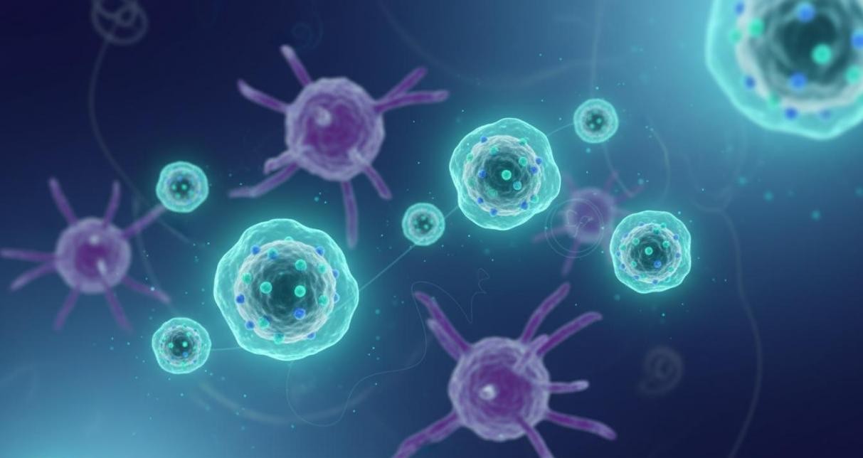 Figure 5: Membrane-coated biomimetic nanoparticles evading macrophage recognition through CD47-mediated "don't eat me" signaling
Demonstrating that membrane-coated nanostructures retain the biological functions of their source cells is a critical validation step. Our in vitro functional testing program confirms immune evasion, targeting specificity, cellular uptake mechanisms, and therapeutic efficacy before advancing to animal studies.
Biological validation capabilities:
- Immune evasion & macrophage uptake: Flow cytometry and confocal microscopy quantifying reduced phagocytosis by RAW 264.7 macrophages compared to uncoated counterparts; CD47 blocking experiments to confirm mechanism.
- Homotypic targeting verification: Competitive binding assays and fluorescence imaging demonstrating preferential adhesion of cancer cell membrane-coated NPs to homologous tumor cells versus heterologous controls.
- Circulation half-life estimation: In vitro serum stability and protein corona analysis predicting in vivo pharmacokinetic behavior.
- Cytotoxicity & biocompatibility: MTT, LDH, and hemolysis assays confirming membrane-coated nanostructures are non-toxic at therapeutic doses.
- Therapeutic potency: Cell viability, apoptosis, and gene expression assays to quantify the biological effect of delivered payloads.
Application-Focused Development Programs
Membrane-coated biomimetic nanostructures have demonstrated transformative potential across multiple therapeutic domains. We structure our development programs around the specific application, assembling the optimal combination of membrane source, nanocore material, payload, and surface engineering to maximize therapeutic impact.
- Oncology & cancer immunotherapy: Homotypic-targeting cancer cell membrane-coated systems for chemotherapy, photothermal/photodynamic therapy, and tumor vaccine applications; combination immunotherapy with checkpoint inhibitors.
- Cardiovascular disease: Platelet membrane-coated nanostructures targeting atherosclerotic plaques, thrombosis, and vascular injury; anti-inflammatory and regenerative payloads.
- Inflammatory & autoimmune conditions: Neutrophil and macrophage membrane-coated systems that home to inflamed tissues for targeted anti-inflammatory drug delivery.
- Infectious disease: Neutrophil and RBC membrane-coated nanostructures for targeted antibiotic delivery and bacterial toxin neutralization.
- Vaccine development: Cancer cell membrane and DC membrane-coated adjuvant nanoparticles for potent antigen-specific immune activation.
Harness the Power of Cellular Mimicry for Your Therapeutic Program
Connect with Eata Nanomaterials to explore how custom membrane-coated biomimetic nanostructures can advance your drug delivery or immunotherapy project. Our scientists will work with you to design a tailored development program aligned with your specific target indication and preclinical milestones.